2Universidade Federal de São Paulo (UNIFESP). São Paulo, Brasil.
3Departamento de Cirugía, Universidade Federal do Ceará (UFC). Ceará, Brasil.
4Hospital das Clínicas, Faculdade de Medicina da Universidade de São Paulo (HCFMUSP). São Paulo, Brasil.
5Ortoclínica, Hospital de Ortopedia. Alagoas, Brasil.
ABSTRACT
Introduction: intra-articular injections are widely used for knee osteoarthritis, yet the optimal strategy for sustained symptom relief remains uncertain.
Objectives: to compare the clinical outcomes of PRP plasma gel, hyaluronic acid, and corticosteroid intraarticular injections, used alone or in combination, for the treatment of knee osteoarthritis.
Methods: this prospective, randomized, single-blind clinical trial included patients with Kellgren–Lawrence grade II–III knee osteoarthritis allocated into 4 treatment groups. Clinical outcomes were assessed using validated patient-reported measures over a 6-month follow-up period.
Results: all groups demonstrated early improvement in pain and function. At 6 months, treatments incorporating PRP plasma gel showed more sustained pain and functional benefits compared with corticosteroid alone. The combination of PRP plasma gel, hyaluronic acid, and corticosteroid yielded the most consistent clinical outcomes.
Conclusion: the combination of PRP plasma gel, hyaluronic acid, and corticosteroid demonstrated more consistent and sustained clinical improvement at 6 months compared with corticosteroid alone, although no statistically significant differences between groups were observed after adjustment. PRP plasma gel alone showed clinical outcomes comparable to hyaluronic acid.
Keywords: Knee osteoarthritis; Platelet-rich plasma; Plasma gel; Hyaluronic acid; Intra-articular injection
Level of evidence: I. Single-blind Prospective Randomized Clinical Trial
PRP plasma gel versus ácido hialurónico versus corticoide para la osteoartrosis de rodilla: ensayo clínico prospectivo y aleatorizado
RESUMEN
Introducción: las infiltraciones intraarticulares son ampliamente utilizadas en el tratamiento de la osteoartrosis de rodilla, aunque persiste controversia sobre la estrategia más eficaz y duradera.
Objetivos: comparar los resultados clínicos del gel de plasma rico en plaquetas (PRP), el ácido hialurónico y el corticosteroide intraarticular, utilizados de forma aislada o combinada, en pacientes con osteoartrosis de rodilla.
Resultados: todos los grupos presentaron mejoría clínica temprana. A los 6 meses, los tratamientos que incluyeron gel de PRP mostraron beneficios más sostenidos en dolor y función en comparación con el corticosteroide aislado. La combinación de gel de PRP, ácido hialurónico y corticosteroide presentó los resultados más consistentes.
Conclusión: la combinación de gel de PRP, ácido hialurónico y corticosteroide mostró una mejoría clínica consistente y sostenida a los 6 meses en comparación con el corticoide aislado, aunque no se observaron diferencias estadísticamente significativas entre los grupos tras el ajuste. El gel de PRP utilizado de forma aislada mostró resultados clínicos comparables al ácido hialurónico.
Palabras clave: osteoartrosis de rodilla; plasma rico en plaquetas; gel de PRP; ácido hialurónico; infiltración intraarticular
Nivel de evidencia: I. Ensayo clínico prospectivo, aleatorizado y simple ciego
INTRODUCCIÓN
Knee osteoarthritis (OA) is a chronic degenerative joint disease characterized by progressive cartilage loss, subchondral bone alterations, and secondary synovial inflammation. These pathological changes result in pain, functional impairment, and diminished quality of life. Leading cause of musculoskeletal disability globally, with its prevalence rising in aging populations and imposing a significant socioeconomic burden.1-3
Intra-articular injections play a central role in the nonoperative management of knee OA.
Corticosteroids are widely used due to their rapid analgesic effect, although their benefits tend to be short-lived. Hyaluronic acid (HA) has been employed as a viscosupplementation strategy, with variable clinical results, particularly in patients with mild to moderate disease. Despite their widespread use, there is no clear consensus regarding the optimal intra-articular therapy for sustained symptom relief.4
Orthobiologic therapies, particularly plateletrich plasma (PRP), have emerged as promising alternatives for knee OA treatment. PRP contains a high concentration of platelets and bioactive growth factors that modulate inflammation and promote tissue repair. Recent studies and meta-analyses have demonstrated superior mid-term clinical outcomes with PRP compared with HA and corticosteroids.5-7
PRP plasma gel represents an advancement over conventional PRP by combining a thermally induced albumin gel matrix with fresh PRP, which allows for prolonged intra-articular retention and sustained release of growth factors..8-11
The objective of this prospective randomized clinical trial was to compare the clinical outcomes of PRP plasma gel, hyaluronic acid, and corticosteroid intraarticular injections, used alone or in combination, for the treatment of knee osteoarthritis.
MATERIALES Y MÉTODOS
Study designThis study was structured as a prospective, randomized, controlled, single-blind clinical trial. Its objective was to compare the clinical effectiveness of various intra-articular injection strategies for treating knee osteoarthritis.
Participants were randomly allocated into four parallel treatment groups and followed longitudinally.
The study was conducted in accordance with the Consolidated Standards of Reporting Trials (CONSORT) guidelines.
The study protocol was reviewed and approved by the Institutional Research Ethics Committee, in compliance with the Declaration of Helsinki and national regulations for research involving human subjects. Written informed consent was obtained from all participants prior to enrollment.
Patient recruitment was conducted between February and April 2025, and all participants were followed for 6 months after the intervention.
ParticipantsEligible patients were adults aged 30 to 90 years with both a clinical and a radiographic diagnosis of knee osteoarthritis, classified as Kellgren–Lawrence grade12 II or III, who were consecutively recruited from the outpatient clinic.
Patients were included if they reported persistent knee pain despite previous conservative treatment and were able to understand and complete all patientreported outcome measures used in the study.
Exclusion criteria comprised the presence of inflammatory rheumatic disease, use of oral, intravenous, or intra-articular corticosteroids within the 12 months preceding enrollment, hemoglobin levels below 11 g/dL, platelet counts lower than 150,000/mm³, known coagulation disorders, active infectious or inflammatory processes near the injection site, and inability to complete the scheduled follow-up assessments.
Randomization and BlindingParticipants were randomly assigned in a 1:1:1:1 ratio to one of four treatment groups using a computergenerated randomization sequence. Allocation concealment was maintained through the use of sealed opaque envelopes.
To maintain participant blinding, blood samples were collected from all enrolled patients, regardless of group assignment. Only participants assigned to PRP-containing groups underwent processing and administration of the hemoderivative. During the injection procedure, a visual barrier was used to prevent participants from seeing the syringe contents, thereby ensuring single-blind conditions.
Intervention groupsParticipants were allocated to one of the following groups:
- Group A: PRP plasma gel + Hyaluronic acid + Corticosteroid
- Group B: Hyaluronic acid + Corticosteroid.
- Group C: PRP plasma gel + Corticosteroid.
- Group D: Corticosteroid alone (control group).
All injections were performed as a single intraarticular procedure.
Preparation of PRP plasma gelSApproximately 20 mL of peripheral venous blood was collected from each participant using two sterile 10-mL plastic vacuum tubes without additives.
Sodium heparin (0.125 mL; 5,000 IU/mL) was added to each tube to prevent coagulation. Samples were centrifuged at 800 × g for 15 minutes using a laboratory centrifuge. After centrifugation, the lower plasma fraction enriched with platelets and the buffy coat layer (approximately 2.5–3.0 mL per tube) were carefully aspirated using sterile syringes and reserved for further processing.
The collected PRP was redistributed using a three way Luer-lock connector into two syringes at a 3:1 ratio. One syringe containing approximately 3.75 mL of PRP was subjected to controlled thermal processing in a laboratory incubator at 75 °C for 15 minutes. This process induced protein denaturation, resulting in a semi-solid albumin gel matrix. The remaining syringe containing approximately 1.25 mL of non-heated PRP was preserved for subsequent mixing.
Following thermal processing, the albumin gel was mechanically mixed with the preserved non-heated PRP using a three-way connector, maintaining a final ratio of 3 parts albumin gel to 1 part fresh PRP. The contents were transferred back and forth between syringes until a homogeneous, stable PRP plasma gel was obtained, yielding a final volume of approximately 5 mL. This final product combined the structural properties of the albumin gel with the biological activity of fresh PRP, allowing sustained intra-articular release of growth factors (Figs. 1 and 2).
Injection procedureTAll intra-articular injections were performed under sterile conditions by experienced orthopedic surgeons. Patients were positioned supine with the knee slightly flexed. Skin antisepsis was performed using chlorhexidine-based solutions, and sterile draping was applied. Local anesthesia was achieved with 2 mL of 2% lidocaine infiltrated into the subcutaneous and pericapsular tissues. A lateral suprapatellar approach was used in all cases.
Prior to injection, therapeutic arthrocentesis was performed to remove any joint effusion when present. The assigned injectate was then administered intra-articularly using a single needle, followed —when applicable— by hyaluronic acid and/or corticosteroid (triamcinolone hexacetonide, 20 mg/mL), according to group allocation. A sterile dressing was applied after the needle was removed. When combination therapies were used, the components were administered sequentially through the same intra-articular access. PRP plasma gel was injected first, followed by hyaluronic acid and finally corticosteroid, without prior mixing in the same syringe.
Post-procedure careParticipants were advised to avoid strenuous physical activity involving the treated knee for at least 24 hours. Cryotherapy was recommended to reduce post-procedural discomfort. Nonsteroidal anti-inflammatory drugs were discouraged during the follow-up period. Opioid analgesics were allowed as rescue medication for a maximum of five days if necessary.
No standardized rehabilitation protocol was prescribed. All patients received the same general post-procedure recommendations, including relative rest for 24 hours and gradual return to activities as tolerated.
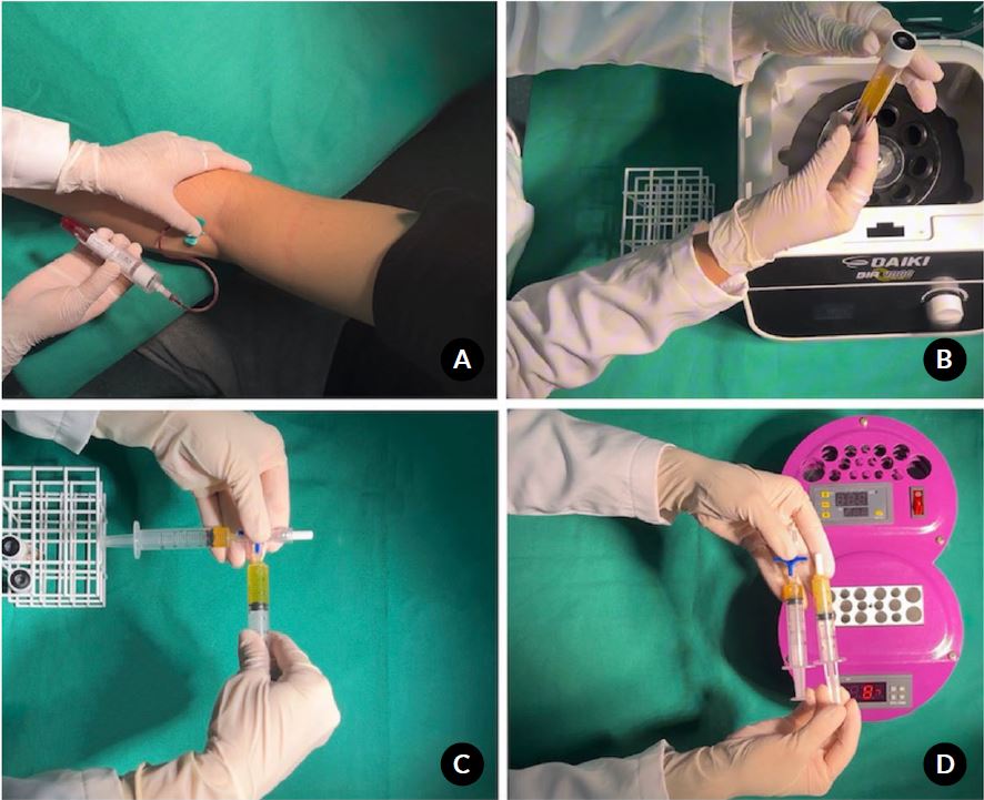
Figure 1. Preparation of plasma gel. A) Peripheral venous blood collection into sterile vacuum tubes. Placement of tubes in the centrifuge, highlighting the importance of proper balance: tubes are positioned diametrically opposite and filled with equal volumes to ensure stability during centrifugation. B) Appearance of the centrifuged blood, showing separation of red blood cells, buffy coat, and plasma. C) Redistribution of PRP using a three-way Luer Lock connector in a 3:1 ratio. Approximately 3.75 mL of PRP is placed in one syringe, while 1.25 mL is kept in another for later use. D) The larger PRP fraction is heated in an incubator at 75 °C for 15 minutes, causing denaturation of albumin, fibrinogen, and other plasma proteins, and forming an albumin-based semisolid gel. The reserved fresh PRP (≈1.25 mL) and the albumin gel (≈3.75 mL) prepared for combination.
Clinical outcomes were assessed using validated patient-reported outcome measures: Visual Analog Scale13 (VAS) for pain, WOMAC,14-16KOOS,17 Pain Catastrophizing Scale18 (PCS), PROMIS Fatigue,19 Global Rating of Change20 (GROC), and Marx Activity Rating Scale.21 Assessments were performed at baseline and at 1, 3, and 6 months after the intervention.
All post-intervention clinical assessments were performed by independent evaluators who were not involved in patient treatment.
Statistical analysisAll clinical and functional data were organized in electronic spreadsheets and analyzed using R software (Mac OS X GUI, version 1.73). Statistical significance was defined as a two-tailed p-value <0.05, at a 95% confidence level.
Categorical variables were summarized as absolute and relative frequencies and compared among groups using Pearson’s Chi-square test or Fisher’s exact test, as appropriate. Continuous variables were assessed for normality using the Shapiro–Wilk test. Normally distributed data were expressed as mean ± standard deviation and compared between groups using oneway analysis of variance (ANOVA) with Tukey post hoc tests, while non-normally distributed data were expressed as median and interquartile range and analyzed using the Kruskal–Wallis test with Dunn’s correction.
Within-group comparisons across time points were performed using paired t-tests for parametric data or Wilcoxon signed-rank tests for nonparametric data. When appropriate, mean changes (delta values) between time points were calculated to assess treatment-related effects over time, and results were graphically presented using mean values with standard deviation error bars. No formal sample size calculation or power analysis was performed.
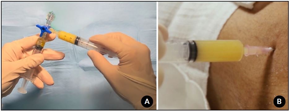
Figure 2. PRP plasma gel combination and application. A) The albumin gel (≈3.75 mL) is mixed with the reserved fresh PRP (≈1.25 mL) using the three-way connector. Multiple passes between syringes ensure homogenization, resulting in approximately 5 mL of PRP plasma gel. B) The PRP plasma gel is transferred into a sterile syringe and injected intra-articularly under aseptic conditions, typically through a suprapatellar or anterolateral portal of the knee.
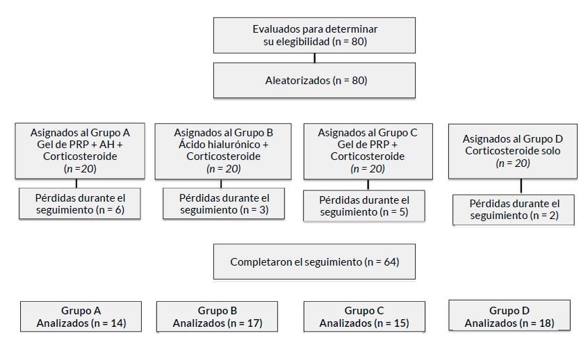
Figure 3. CONSORT flow diagram illustrating patient enrollment, randomization, allocation to treatment groups, follow-up, and final analysis in this prospective randomized clinical trial.
RESULTS
A total of 80 patients were initially enrolled and randomized; 64 completed follow-up and were included in the final analysis (Fig. 3). The overall mean age was 62.1 ± 8.9 years (range, 42–78), and 71% of participants were female. All patients had radiographic knee osteoarthritis classified as Kellgren–Lawrence grade II–III. Participant blinding was maintained throughout the trial using the predefined single-blind protocol. Final group distribution among participants with complete follow-up was Group A (PRP plasma gel + hyaluronic acid + corticosteroid), n = 14; Group B (hyaluronic acid + corticosteroid), n = 17; Group C (PRP plasma gel + corticosteroid), n = 15; and Group D (corticosteroid alone), n = 18. Outcomes were assessed longitudinally from baseline through follow-up time points as defined in the protocol. No major or minor complications, including infection, exacerbation of persistent pain, or adverse reactions, were observed during the study period.
At the 6-month follow-up, all treatment groups demonstrated a significant reduction in pain intensity compared with baseline at early time points (Fig. 4 and Table 1). At 1 and 3 months, Groups A, B, C, and D showed statistically significant decreases in VAS scores (p<0.05 for all). However, at 6 months, sustained pain reduction remained statistically significant only in Groups A (PRP plasma gel + hyaluronic acid + corticosteroid; p = 0.0037), B (hyaluronic acid + corticosteroid; p = 0.0017), and C (PRP plasma gel + corticosteroid; p = 0.0010), whereas the corticosteroid-only group (Group D) no longer demonstrated a significant difference from baseline (p = 0.1878).
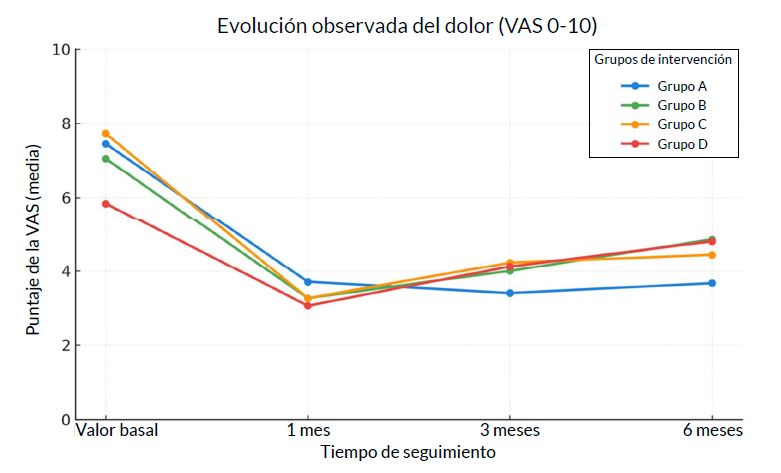
Figure 4. Visual Analog Scale (VAS) pain scores at baseline, 1, 3, and 6 months following intra-articular treatment. PRP plasma gel–based groups demonstrated sustained pain reduction at 6 months, whereas the corticosteroid-only group showed loss of analgesic effect over time.
| Group | Intervention | Baseline | 1 month | 3 months | 6 months | p (Baseline vs. 1M) | p (Baseline vs. 3M) | p (Baseline vs. 6M) |
|---|---|---|---|---|---|---|---|---|
| A | PRP plasma gel + HA + Corticosteroid | 7.47 ± 2.10 | 4.00 ± 3.32 | 3.88 ± 2.75 | 4.40 ± 3.17 | 0.0003 | 0.0436 | 0.0037 |
| B | HA + Corticosteroid | 6.65 ± 1.90 | 3.46 ± 2.22 | 4.06 ± 2.17 | 5.00 ± 2.48 | 0.0003 | 0.0003 | 0.0017 |
| C | PRP plasma gel + Corticosteroid | 7.79 ± 2.26 | 3.47 ± 2.88 | 4.21 ± 2.83 | 4.50 ± 3.03 | 0.0001 | 0.0002 | 0.0010 |
| D | Corticosteroid only | 5.56 ± 2.68 | 2.80 ± 3.23 | 4.07 ± 3.63 | 4.76 ± 3.53 | 0.0050 | 0.0456 | 0.1878 |
At the 6-month follow-up, Groups A, B, and C exhibited significant within-group improvements in the WOMAC pain and function domains compared with baseline (p <0.05 for all) (Fig. 5). In contrast, Group D did not show significant improvement in WOMAC pain (p = 0.115) or function (p = 0.255) at 6 months. Significant improvement in WOMAC stiffness scores was observed only in Group A (p <0.05), whereas Groups B, C, and D did not exhibit statistically significant changes in stiffness.
At 6 months, KOOS subscale analysis indicated a consistent trend favoring Group A across pain, symptoms, activities of daily living, sport/ recreation function, and knee-related quality of life.
Nevertheless, after adjustment for baseline values using ANCOVA, no statistically significant differences between groups were identified at the 6-month time point (Type III ANCOVA, p >0.05 for all domains).
Significant reductions in PCS scores from baseline to 6 months were observed in Group A (Δ = −13.42; p = 0.0043), Group B (Δ = −13.47; p = 0.0040), and Group C (Δ = −13.50; p = 0.0202), indicating meaningful improvement in pain-related catastrophizing.
Group D showed a smaller non-significant reduction (Δ = −8.38; p = 0.0843). Adjusted between-group comparisons at 6 months were not statistically significant (ANCOVA p = 0.760).
PROMIS Fatigue scores improved over time in all treatment, but only Group A demonstrated a statistically significant within-group improvement at 6 months (Δ = −16.83; p = 0.0395). No statistically significant between-group differences were detected after adjustment for baseline values (ANCOVA p = 0.2944).
At the 6-month evaluation, patient-perceived improvement assessed by the GROC scale showed significant within-group improvement across all treatment arms. The greatest magnitude of perceived improvement was observed in Group A (Δ = +5.14; p = 0.0135) and Group C (Δ = +4.67; p = 0.0132).
Adjusted between-group comparisons did not reveal statistically significant differences at 6 months (ANCOVA p = 0.953).
MARX activity scores demonstrated a nonsignificant upward trend from baseline to 6 months in all groups: Group A (Δ = +1.33; p = 0.1661), Group B (Δ = +0.71; p = 0.3061), Group C (Δ = +0.58; p = 0.3934), and Group D (Δ = +1.44; p = 0.1216). No statistically significant differences within-group or between-groups were identified at the 6-month follow-up (ANCOVA p = 0.733).
DISCUSSION
The primary finding of this study was that intraarticular strategies incorporating PRP plasma gel were associated with more sustained clinical improvement over time compared with corticosteroid alone. However, no statistically significant differences between groups were observed after adjustment.
Although all treatment arms demonstrated early pain relief, only the PRP-containing groups maintained improvements over time, whereas the corticosteroidonly group showed loss of efficacy at mid-term followup. Despite the absence of statistically significant differences between groups after adjustment, the observed sustained improvements in pain and function may be clinically meaningful, particularly in the context of chronic conditions such as knee osteoarthritis, where long-term symptom control is a key therapeutic goal.
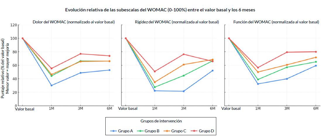
Figure 5. WOMAC subscale scores (pain, stiffness, and function) from baseline to 6-month follow-up. Significant improvements in pain and function were observed in PRP plasma gel–containing groups, with stiffness improvement occurring exclusively in the combined PRP plasma gel, hyaluronic acid, and corticosteroid group.
This temporal pattern is consistent with previous literature. Corticosteroids are known to provide rapid analgesia with limited durability, a finding repeatedly reported in clinical trials and systematic reviews. In contrast, biologically active therapies such as PRP have demonstrated superior mid-term outcomes when compared with corticosteroids and hyaluronic acid, particularly for pain and function. Meta-analyses by Belk et al.22 and Khalid et al.23 have shown that PRP results in greater improvements in WOMAC and VAS scores at follow-ups beyond 3 months, supporting the sustained analgesic effect observed in the present study
Functional outcomes assessed by WOMAC further support these findings. At 6 months, patients who received with PRP plasma gel —with or without hyaluronic acid— demonstrated significant improvements in pain and function, whereas corticosteroid alone failed to produce meaningful functional gains. Notably, improvement in WOMAC stiffness was observed only in the group that received the combined PRP plasma gel, hyaluronic acid, and corticosteroid injection, suggesting a potential synergistic effect.23 Similar benefits of combined PRP and hyaluronic acid therapies have been reported in recent systematic reviews, including the metaanalysis by Karasavvidis et al.,24 which demonstrated superior functional outcomes compared with hyaluronic acid alone.
Although KOOS outcomes did not reach statistical significance after adjustment for between-group differences, the consistent trend favoring the combined PRP plasma gel strategy across all subdomains suggests a clinically relevant effect that may not have been fully captured due to sample size limitations. Previous studies evaluating PRP in knee osteoarthritis have similarly reported clinically meaningful improvements without consistent statistical separation across all KOOS subscales, particularly in heterogeneous patient populations.22,23
Psychosocial and patient-centered outcomes provide additional evidence supporting the effectiveness of sustained biologic modulation.
Significant reductions in pain catastrophizing were observed only in groups receiving PRP and/or hyaluronic acid, which is consistent with evidence that longer-lasting symptom control improves pain perception and coping strategies.18,22 Furthermore, only the combined PRP plasma gel group exhibited significant improvement in PROMIS Fatigue scores, suggesting broader functional and quality-of-life benefits beyond pain relief.19 These results align with previous reports suggesting that biologic intraarticular therapies may modulate inflammatory mediators and synovial biomarkers associated with systemic fatigue and symptom burden.25,26
The sustained performance of PRP plasma gel can be attributed to its unique structural characteristics.
Unlike conventional liquid PRP, plasma gel contains a thermally induced albumin matrix combined with fresh PRP, which enables prolonged intra-articular retention and gradual release of growth factors such as PDGF, TGF-β, and VEGF.8-11 Experimental and translational studies, including those by Godoi et al.11 and Nakamura et al.,8 have shown that plasma-based gels serve as effective biological carriers, facilitating controlled release and prolonged bioactivity, which may explain the sustained clinical effects observed in this trial.
Although corticosteroids have been rshown to inhibit specific growth factors, their inclusion in this study was informed by both clinical and methodological considerations. Clinically, corticosteroids provide rapid analgesic and anti-inflammatory effects, which may contribute to early symptom relief while the biological effects of PRP plasma gel take time to emerge, reflecting a pragmatic real-world strategy.
In addition, corticosteroids were administered at a low dose, as lower doses have been associated with reduced deleterious effects on mesenchymal cells.
Methodologically, the use of corticosteroids across all treatment groups allowed for a more controlled comparison, helping to isolate the additional effects of PRP plasma gel and hyaluronic acid beyond the known effects of corticosteroids. This design aimed to reduce confounding and better clarify the contribution of each therapeutic component.
From both clinical and economic perspectives, the comparison between PRP plasma gel plus corticosteroid and hyaluronic acid plus corticosteroid is particularly relevant. Although both strategies demonstrated comparable clinical improvements, PRP plasma gel represents an autologous therapy that can be prepared with standard laboratory equipment, potentially at lower cost than commercially available hyaluronic acid formulations. In resourcelimited healthcare systems, this distinction may have significant implications for accessibility and cost-effectiveness, without compromising clinical outcomes.27
Further research directly comparing PRP plasma gel with conventional PRP formulations is necessary to clarify the potential benefits of this modified biological approach.
This study has limitations, such as a modest sample size, lack of a formal sample size calculation, unequal group sizes following attrition, and a 6-month followup. Additionally, imaging or biochemical markers were not assessed, which could have provided additional mechanistic insights. However, the study’s strengths include its prospective randomized design, the use of multiple validated outcome measures, and direct comparison of clinically relevant intra-articular interventions.
CONCLUSION
The combination of PRP plasma gel, hyaluronic acid, and corticosteroid resulted in more consistent and sustained clinical improvement at 6 months compared to corticosteroid alone. However, no statistically significant differences between groups were observed after adjustment. PRP plasma gel alone showed clinical outcomes comparable to those of hyaluronic acid.
REFERENCES
- Keyes GW, Carr AJ, Miller RK, Goodfellow JW. The radiographic classification of medial gonarthrosis: correlation with operation methods in 200 knees. Acta Orthop Scand. 1992;63(5):497-501
- Kellgren JH, Lawrence JS. Radiological assessment of osteoarthrosis. Ann Rheum Dis. 1957;16(4):494-502
- Ahlbäck S. Osteoarthrosis of the knee: a radiographic investigation. Acta Radiol Diagn (Stockh). 1968:Suppl 277:7-72
- Arliani GG, Durigon TS, Pedroso JP, Ferreira GF, Oksman D, Oliveira VO. Intra-articular infiltration of platelet-rich plasma versus hyaluronic acid in patients with primary knee osteoarthritis: preliminary results from a randomized clinical trial. Rev Bras Ortop (Sao Paulo). 2021;57(3):402-408
- Piuzzi NS, Khlopas A, Newman JM, Ng M, Roche M, Husni ME, et al. Bone marrow cellular therapies: novel therapy for knee osteoarthritis. J Knee Surg. 2018;31(1):22-26
- Keeling LE, Belk JW, Kraeutler MJ, Kallner AC, Lindsay A, McCarty EC, et al. Bone marrow aspirate concentrate for the treatment of knee osteoarthritis: a systematic review. Am J Sports Med. 2022;50(8):2315-2323
- Andrade Cunha PF, da Silva RBB. Knee osteoarthritis and bone marrow aspirate as a treatment choice: narrative review. Res Soc Dev. 2021;10(7)
- Nakamura M, Masuki H, Kawabata H, Watanabe T, Watanabe T, Tsujino T, et al. Plasma gel made of platelet-poor plasma: in vitro verification as a carrier of polyphosphate. Biomedicines. 2023;11(11):2871
- Liu Q, Zhang N, Li Z, He H. Efficacy of autologous platelet-rich plasma gel in the treatment of refractory pressure injuries and its effect on wound healing time and patient quality of life. Clinics (Sao Paulo). 2021;76:e2355
- Wang M, Gao W. Fixation of platelet-rich plasma and fibrin gels on knee cartilage defects after microfracture with arthroscopy. Int Orthop. 2022;46(8):1761-1766
- Godoi TTF, Rodrigues BL, Huber SC, Santana MHA, da Fonseca LF, Santos GS, et al. Platelet-rich plasma gel matrix (PRP-GM): description of a new technique. Bioengineering (Basel). 2022;9(12):817
- Schiphof D, Boers M, Bierma-Zeinstra SM. Differences in descriptions of Kellgren and Lawrence grades of knee osteoarthritis. Ann Rheum Dis. 2008;67(7):1034-1036
- Campolina AG, Bortoluzzo AB, Ferraz MB, Ciconelli RM. Validação da versão brasileira do questionário genérico de qualidade de vida short-form 6 dimensions (SF-6D Brasil) [Validation of the Brazilian version of the generic six-dimensional short form quality of life questionnaire (SF-6D Brazil)]. Cien Saude Colet. 2011;16(7):3103-3110. Portuguese
- Bellamy N, Buchanan WW, Goldsmith CH, Campbell J, Stitt LW. Validation study of WOMAC: a health status instrument for measuring clinically important patient relevant outcomes to antirheumatic drug therapy in patients with osteoarthritis of the hip or knee. J Rheumatol. 1988;15(12):1833-1840
- Oliveira AM, Peccin MS, Silva KN, Teixeira LE, Trevisani VF. Impact of exercise on the functional capacity and pain of patients with knee osteoarthritis: a randomized clinical trial. Rev Bras Reumatol. 2012;52(6):876-882
- Hmamouchi I, Allali F, Tahiri L, Khazzani H, Mansouri LE, Ali Ou Alla S, et al. Clinically important improvement in the WOMAC and predictor factors for response to non-specific non-steroidal antiinflammatory drugs in osteoarthritic patients: a prospective study. BMC Res Notes. 2012;5:58
- Roos EM, Roos HP, Lohmander LS, Ekdahl C, Beynnon BD. Knee Injury and Osteoarthritis Outcome Score (KOOS): development of a self administered outcome measure. J Orthop Sports Phys Ther. 1998;28(2):88-96
- Darnall BD, Sturgeon JA, Cook KF, Taub CJ, Roy A, Burns JW, et al. Development and validation of a daily pain catastrophizing scale. J Pain. 2017;18(9):1139-1149
- Cheung EC, Moore LK, Flores SE, Lansdown DA, Feeley BT, Zhang AL. Correlation of PROMIS with orthopaedic patientreported outcome measures. JBJS Rev. 2019;7(8):e9
- Schmitt J, Abbott JH. Global ratings of change do not accurately reflect functional change over time in clinical practice. J Orthop Sports Phys Ther. 2015;45(2):106-111, D1-D3
- Çil ET, Ebru Akbu a Koç, Akbuga Koc E, Su Arkun H, Ba cıba ı G , et al. Turkish validity and reliability of the Marx Activity Rating Scale (MARS) for patients with knee problems. BAUN Health Sci J. 2024;13(1):60-66
- Belk JW, Kraeutler MJ, Houck DA, Goodrich JA, Dragoo JL, McCarty EC. Platelet-rich plasma versus hyaluronic acid for knee osteoarthritis: a systematic review and meta-analysis of randomized controlled trials. Am J Sports Med. 2021;49(1):249-260
- Khalid S, Ali A, Deepak F, Zulfiqar MS, Malik LU, Fouzan Z, et al. Comparative effectiveness of intra-articular therapies in knee osteoarthritis: a meta-analysis comparing platelet-rich plasma (PRP) with other treatment modalities. Ann Med Surg (Lond). 2023;86(1):361-372
- Karasavvidis T, Totlis T, Gilat R, Cole BJ. Platelet-rich plasma combined with hyaluronic acid improves pain and function compared with hyaluronic acid alone in knee osteoarthritis: a systematic review and meta-analysis. Arthroscopy. 2021;37(4):1277-1287.e1
- Li T, Li Y, Li W, et al. Impact of autologous platelet-rich plasma therapy vs. hyaluronic acid on synovial fluid biomarkers in knee osteoarthritis: a randomized controlled clinical trial. Front Med. 2023;10
- Pretorius J, Habash M, Ghobrial B, Alnajjar R, Ellanti P. Current status and advancements in platelet-rich plasma therapy. Cureus. 2023;15(10):e47176
- Slimi F, Zribi W, Trigui M, Amri R, Gouiaa N, Abid C, et al. The effectiveness of platelet-rich plasma gel on full-thickness cartilage defect repair in a rabbit model. Bone Joint Res. 2021;10(3):192-202